 |
| Antibiotic tests 1-4 |
 |
| Antibiotic tests 5-8 |
The measured diameters of the bacteria surrounding the antibiotics are as follows:
Penicillin: 8 mm (resistant)
Novobiocin: 0 mm (resistant)
Neomycin: 14 mm (intermediate)
Streptomycin: 19 mm (sensitive)
Tetracycline: 16 mm (intermediate)
Erythromycin: 0 mm (resistant)
Amoxicillin: 22 mm (sensitive)
Oregano: 20 mm (sensitive)
 |
| Measuring the diameter |
We also analyzed the selective and differential media plates. We used mannitol salt agar plates to see if Molly had the bacteria Staphylcoccus aureus (MRSA). Using a sterile cotton swab, we dipped the swab into saline solution and swiped it around the inside of Molly's nostrils. We then spread the swab around on the mannitol salt agar plate to cover the entire plate. We incubated the plate at 37°C (the temperature of Molly's body). After 24 hours, we did not find the Staphylococcus aureus bacteria in Molly's nostrils (Thank goodness!)
 |
| Negative!! |
The next experiment was throat swabs! This test s used for detecting the bacteria streptococcus (strep throat). Lindsey and I both had our throats swab because I had a sore throat and Lindsey wasn't sick so we wanted to compare the two. Using a sterile cotton swab, we got bacteria from the throat and spread it around the blood agar plate, covering the entire plate. We incubated the plate at 37°C (the temperature of the human body). Both plates turned out to be α lysis which is a particle sensitivity to hemolysis. However my half of the plate showed a spot that could be the streptococcus bacteria!
 |
| Our throat swabs! |
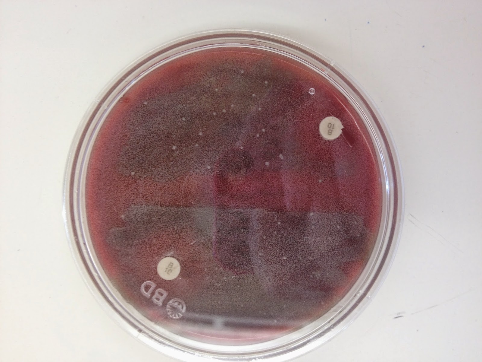
The final four selective and differential plates were the blood agar plate, MacConkey agar plate, Phenyethyl alcohol agar plate, and EMO plate. On each of the different plates, using the aseptic technique, we drew a squiggle line and incubated the plate at 37°C overnight.
The blood agar plate showed α partial hemolysis.
 |
| Our results are on the left |
 |
| Our alpha results on the right |
The MacConkey Agar was positive because there was no color change. Gram-negative bacteria grows.
Phenyethyl alcohol agar plate was negative because only gram-positive bacteria grows and our bacteria is gram-negative.
 |
| Our bacteria on the right- no growth |
Our EMO plate was positive as there was clearly growth!
 |
| Look at that thing! |
 |
The next experiment was the ELISA test. The ELISA test demonstrates the use of antigens and antibodies. Antigens produce antibodies to kill substances foreign to the body. The test was used to see if the HIV virus was present in different samples. First, we labeled each of the 12 wells properly. Then we added 50 microliters of the purified agent into each well and let that sit for 5 minutes so the antigen can bind to the well. After 5 minutes, we poured the solution onto a paper towel and washed the wells with a wash buffer. Now, it was time to add the antibody. We then added 50 microliters of the positive control to the first 3 wells, 50 microliters of the negative control to the next 3 wells. We then added 50 microliters of the first sample, 20, into the wells 7-9 and the 50 microliters of the second sample, 43, to wells 10-12. After 5 minutes, we washed the wells with the wash buffer. We then added 50 microliters of the second antibody to all 12 wells. After 5 minutes, we washed the wells with the wash buffer twice. Finally, was added 50 microliters of the enzyme substrate to the 12 wells. After 5 minutes, we observed the sample 43 was HIV positive.
 |
| ELISA test |
 |
| Our materials |
 |
| Positive patient 43! |
No comments:
Post a Comment