After final exams were done, 8 students decided the semester wasn't long enough and decided to take on one more class. We entered the laboratory and Dr. Pathakamuri gave a small introduction to the class. He told us how this class is important to nursing majors and the health science relations, in general. He said that patients will have germs and illnesses and we, as professionals, need to be able to aid in the treatment of the illnesses quickly. We need to be able to properly identify the bacteria to be able to treat it efficiently. Also in our introduction, Dr. Pathakamuri gave a brief introduction about the laboratory safety. Once we enter the lab, we need to put on our lab coats, wash our hands thoroughly, and clean our bench area with disinfectant to get rid of any bacteria. When we left the lab, we need to clean our bench area, wash our hands, and hang up our lab coats.

To really see and understand how clean our hands were after they were washed, we did an experiment. Lindsey and I received a Petri dish with a culture medium. We divided our dish into four sections using a China marker. We pressed our thumbs into the medium and quickly covered the dish to prevent any contamination. Then we washed our hands, we pressed our thumbs again into the medium. We covered the dish and put them in a 37
°C incubator. This allowed the bacteria to grow overnight.
 |
| Our Petri Dish with our thumb prints before and after washing |
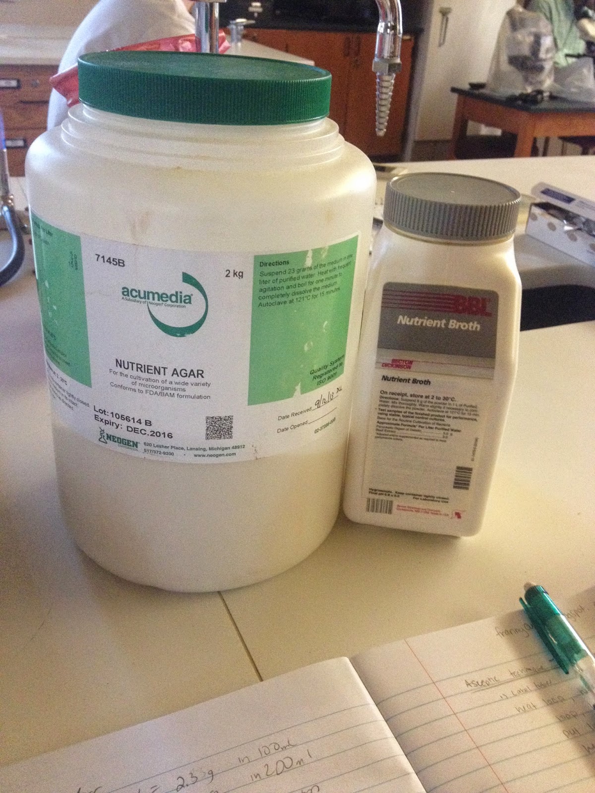
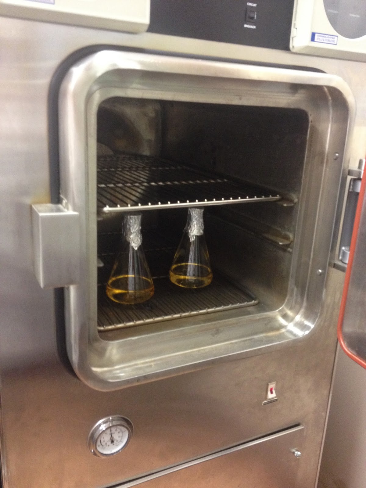
Next, we made our own media culture from Nutrient Agar or Nutrient Broth. Our group was assigned Nutrient Agar. We measured 4.6 grams of Nutrient Agar and added it to a beaker of 200 mL of distilled water and covered the beaker with foil to prevent water loss. The mixture was placed in the autoclave for 20 minutes under 15 lbs of pressure at 121
°C. The autoclave uses moist heat for sterilization. Once the mixture cooled to approximately 30
°C, the mixture was poured into Petri dishes to cover the bottom. The dish was covered to prevent contamination and the liquid had turned to gel within minutes. The dishes were placed in a 24
°C incubator.
 |
| Weighing our Nutrient Agar |
 |
| Placing our mixture in the autoclave |
 |
| Removing our mixture from the autoclave |
 |
| Gel media cultures |
We learned about Aseptic techniques to transport bacteria from one tube to another under sterile conditions. We lit the Bunsen burner and were given two test tubes of liquid. The inoculation loop was sterilized over the Bunsen burner, the test tube was slightly heated to remove bacteria, the inoculation loop was placed in the test tube to get a thin film covering the loop, and the tube was heated and closed. The second test tube was heated slightly and the bacteria was placed in the second tube. The second tube was heated and closed. The inoculation loop was heated a final time for sterilization.
 |
| Sterilizing the inoculation loop |
Our final experiment of the day was to realize how many colonies of bacteria surround you. For this experiment, we decided to swab the silverware water container in the cafeteria. We figured this would have tons of bacteria since this is where all the dirty silverware is placed. We swiped the swab in the dirty water and spread it across the Petri dish. We put the dish in the 24°C incubator (room temperature). We left the bacteria grow over two nights.
We examined the colony morphology of the bacteria. The whole colony form had three types of bacteria: puntiform, circular, and rhizoid. The colony pigmentation was white/colorless and the colony margin was entire. The elevation of the puntiform colony was raised. The elevation of the circular colony was raised. The elevation of the rhizoid colony was undulate.
 |
| Silverware Bin Bacteria |

No comments:
Post a Comment